OCT angiography plays a key role in the early stages of glaucoma
Author: Dr. Maria Benova
The method is particularly useful for distinguishing changes related to myopia and glaucoma.
The examination is the method of choice for diagnosing and monitoring glaucoma in myopic eyes.
Last week, we marked World Glaucoma Week (At the eye clinic ‘PENTAGRAM’ we offered free check-ups on this occasion). The initiative aims to raise awareness about the disease and encourage its early detection through regular preventive check-ups. The term glaucoma encompasses a number of nosological entities with different and not fully understood causes and development mechanisms. What is common for this group of eye diseases is that they lead to damage to the optic nerve and cause irreversible vision loss, if left untreated.
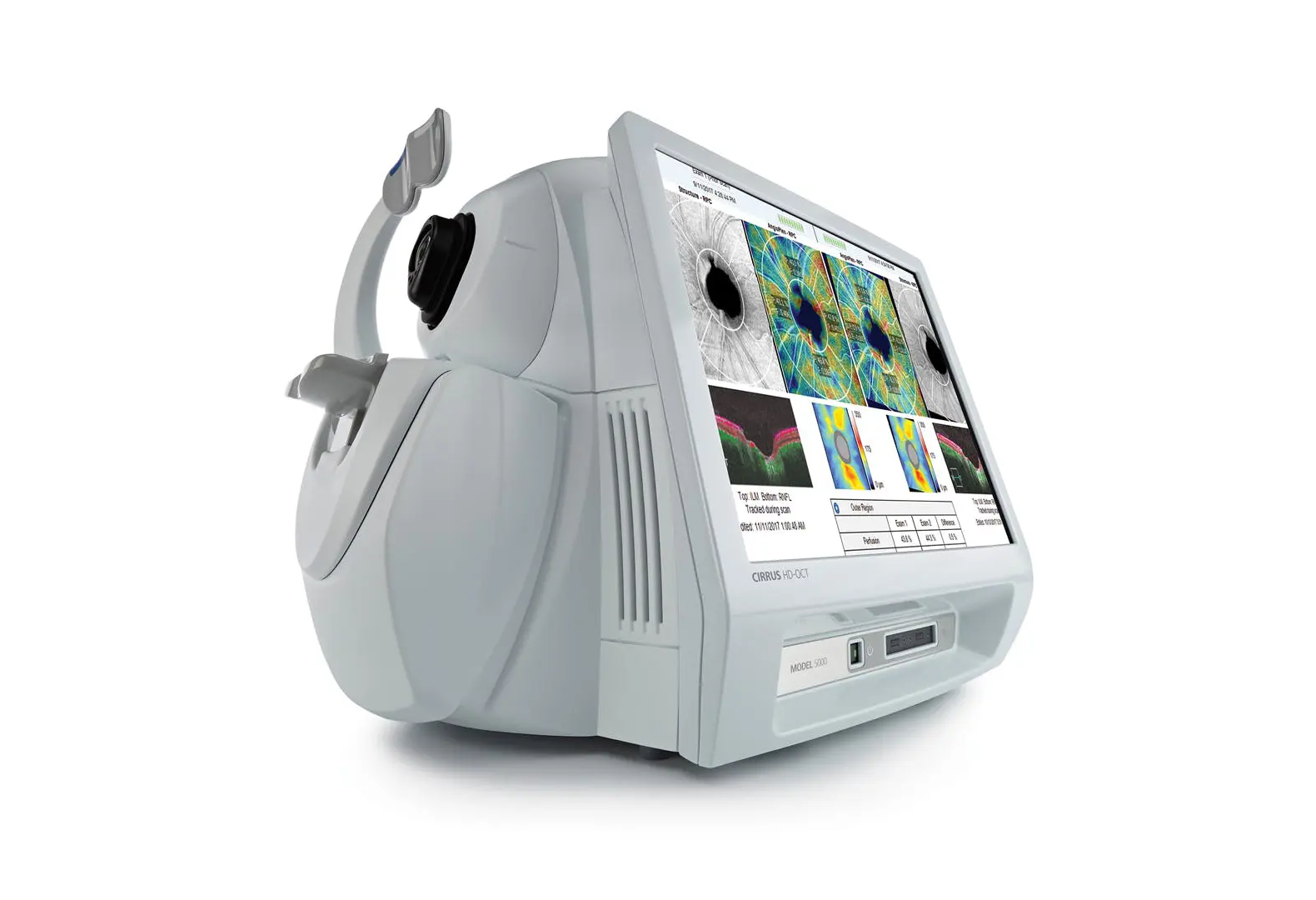
Dr. Maria Benova explains the role of OCT angiography – a high-tech examination performed at the ‘PENTAGRAM’ Eye Clinic and all major eye centers for diagnosing glaucoma and monitoring the disease. Dr. Maria Benova. Dr. Benova is an ophthalmology specialist with over 20 years of experience and professional interests in the field of diagnosing and treating diseases of the anterior eye segment, glaucoma, cataracts, as well as medical retina and oculoplastics. At MTC ‘PENTAGRAM’, she consults patients with referrals from the NHIF, through a health fund, or for a fee. Appointments can be made online via HERE or by calling 0700 91 191.
Primary open-angle glaucoma is the most common type of glaucoma, characterized by an asymptomatic onset and late diagnosis. It is called the ‘silent thief of sight’ because vision loss typically occurs slowly and asymptomatically over a long period of time..
One of the main risk factors for the development of glaucoma is elevated intraocular pressure, which leads to damage to the nerve fibers that form the optic nerve and to the death of retinal ganglion cells. Nonetheless, a significant portion of patients develop primary open-angle glaucoma with intraocular pressure values within the normal range.
This is why one of the major efforts in modern ophthalmology is focused on identifying risk factors and detecting the disease early. Especially when it is suspected that risk factors other than intraocular pressure, play a relatively greater role in primary open-angle glaucoma.
Fortunately, we have many technologies, to assist us in diagnosing glaucoma. However, new advancements are always welcome, especially if they enhance our ability to detect and monitor the disease while also making clinic visits more comfortable for patients.
OCT angiography is just such a type of technology. It allows monitoring of the blood flow in the retina, providing us with a new way to assess the condition of ganglion cells, which may be destroyed by the disease.
One of the theories about the development of glaucoma is the vascular theory, according to which glaucomatous optic neuropathy results from insufficient blood flow to the optic nerve head due to elevated IOP or other factors reducing blood supply. Glaucomatous damage to the optic nerve occurs under conditions of reduced ocular perfusion pressure. For this reason, glaucoma is often associated with diseases involving vascular dysregulation – for example migraine и Raynaud’s syndrome. Systemic arterial hypertension is associated with the occurrence of local vascular dysregulation and can also be considered a risk factor for glaucoma. The results of studies in this direction do not provide a definitive answer.
Interestingly, when OCT angiography was initially used in glaucoma, reduced vascular density was found in the lamina cribrosa plane. The lamina cribrosa is a lattice, sieve-like structure located at the back of the eye, where the bundles of nerve fibers of the optic nerve exit the eyeball to head towards the brain. The blood flow deficit in the lamina cribrosa is intriguing because this is the potential site where increased intraocular pressure may affect the perfusion of the optic nerve. However, for routine diagnostic purposes, the optic nerve head is not an ideal imaging target due to the overshadowing of neural tissue by several large blood vessels and the highly variable geometry of the optic disc’s edge. It turns out that the superficial vascular zone is the most suitable for glaucoma examination. OCT angiographic scans are performed in the macula and optic nerve area.
It has been found that the reproducibility of blood flow measurements is poor. The main reason is that the speed and volume of blood flow are influenced by variations in the patient’s physiological state, the oxygen concentration in the inhaled gas mixture, and visual stimulation. There are probably many other factors that affect blood flow. The reproducibility of vessel density measurements via OCT angiography, however, is excellent. For glaucoma evaluation, vascular density and, in particular, capillary density is the ideal indicator for diagnosis and monitoring.
For all glaucoma patients, we now routinely perform OCT angiography. But in several clinical scenarios, OCTA provides us with additional informativeness. For example, in very early stages of glaucoma, dysfunctional ganglion cells have reduced metabolism, which leads to reduced capillary density. This reduced density is detected by OCT angiography, before the death and subsequent thinning of the nerve fiber layer and retinal ganglion cells, which can be detected through structural OCT.

In patients with high myopia, the data from standard glaucoma tests such as computer perimetry and conventional OCT are inaccurate, which makes it difficult to distinguish between truedisease-related changes and factors associated with high myopia. On the other hand, vessel density does not decrease in a healthy myopic eye, which is why the method is useful for diagnosing and monitoring glaucoma in myopic eyes.
OCT angiography proves to be useful in patients with glaucoma and the presence of a paracentral defect in the visual field. In this type of change, OCT angiography is more sensitive than standard OCT and detects vascular density deficits before structural OCT changes occur.
Unlike structural OCT, OCT angiography does not exhibit a significant threshold effect and remains a useful method for monitoring glaucoma progression in advanced stages of the disease.
OCT angiography is a useful method for both patients and ophthalmologists in our efforts to better understand the pathogenesis of this socially significant disease. The method can be used as a complement to standard glaucoma tests and is particularly important in patients with high myopia, in very early as well as advanced glaucoma and in some specific changes in the visual field. The limitations of the method are the frequent presence of artifacts in the images and the smaller number of steps in the dynamic range of OCT angiography parameters, so the results must be interpreted with care by a specialist ophthalmologist.

For yet another year, ‘PENTAGRAM’ is part of the ‘CHRISTMAS TREES OF TALENT’ campaign, funding young talents
The 'Pentagram' Eye Clinic supported the 'Christmas Trees of Talent' campaign by the 'Stoyan Kambarev' Foundation to help large families.
Read More
The PreserFlo Implant – A Proven Effective Tool in the Fight Against Glaucoma
Glaucoma – ‘the silent thief of sight’ – is often how this disease is described, as it ‘steals’ vision and.
Read More